
Shoulder Arthroscopy
Arthroscopy is a procedure where orthopaedic surgeons use to inspect, diagnose, and repair problems inside a joint.
It has made diagnosis, treatment, and recovery from surgery easier and faster than was once thought possible. Improvements to shoulder arthroscopy occur every year as new instruments and techniques are developed.
Anatomy
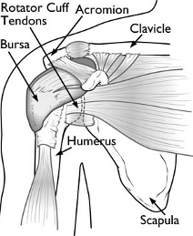
Your shoulder is a complex joint that is capable of more motion than any other joint in your body. It is made up of three bones: your upper arm bone (humerus), your shoulder blade (scapula), and your collarbone (clavicle).
Ball and Socket. The head of your upper arm bone fits into a rounded socket in your shoulder blade. This socket is called the glenoid. A slippery tissue called articular cartilage covers the surface of the ball and the socket. It creates a smooth, frictionless surface that helps the bones glide easily across each other.
The glenoid is ringed by strong fibrous cartilage called the labrum. The labrum forms a gasket around the socket, adds stability, and cushions the joint.
Shoulder Capsule. The joint is surrounded by bands of tissue called ligaments. They form a capsule that holds the joint together. The undersurface of the capsule is lined by a thin membrane called the synovium. It produces synovial fluid that lubricates the shoulder joint.
Rotator Cuff. Four tendons surround the shoulder capsule and help keep your arm bone centered in your shoulder socket. This thick tendon material is called the rotator cuff. The cuff covers the head of the humerus and attaches it to your shoulder blade.
Bursa. There is a lubricating sac called a bursa between the rotator cuff and the bone on top of your shoulder (acromion). The bursa helps the rotator cuff tendons glide smoothly when you move your arm.
When Shoulder Arthroscopy Is Recommended
If you have a painful condition that does not respond to nonsurgical treatment. Nonsurgical treatment includes rest, physical therapy, and medications or injections that can reduce inflammation. Inflammation is one of your body's normal reactions to injury or disease. In an injured or diseased shoulder joint, inflammation causes swelling, pain, and stiffness.
Injury, overuse, and age-related wear and tear are responsible for most shoulder problems. Shoulder arthroscopy may relieve painful symptoms of many problems that damage the rotator cuff tendons, labrum, articular cartilage, and other soft tissues surrounding the joint.
- Rotator cuff repair
- Bone spur removal
- Removal or repair of the labrum
- Repair of ligaments
- Removal of inflamed tissue or loose cartilage
- Repair for recurrent shoulder dislocation
Normal Anatomy of the Shoulder
Shoulder Arthroscopy
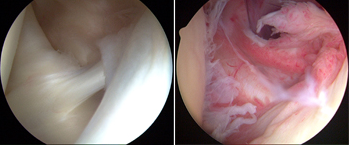
These photos taken through an arthroscope show a normal shoulder joint lining (left) and an inflamed joint lining damaged by frozen shoulder.
What happens during Surgical Procedure?
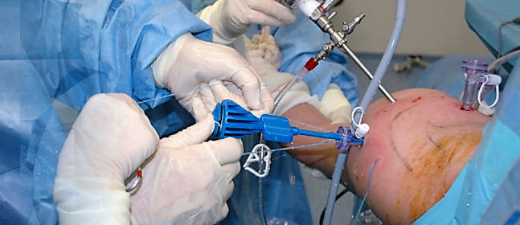
Once in the operating room, you will be positioned so that I can easily adjust the arthroscope to have a clear view of the inside of your shoulder. I personally prefer Lateral decubitius position. In this position the patient lies on his or her side on an operating table.
I will first inject fluid into the shoulder to inflate the joint. This makes it easier to see all the structures of your shoulder through the arthroscope. Then I will make a small puncture in your shoulder (about the size of a buttonhole) for the arthroscope. Fluid flows through the arthroscope to keep the view clear and control any bleeding. Images from the arthroscope are projected on the video screen showing your surgeon the inside of your shoulder and any damage.
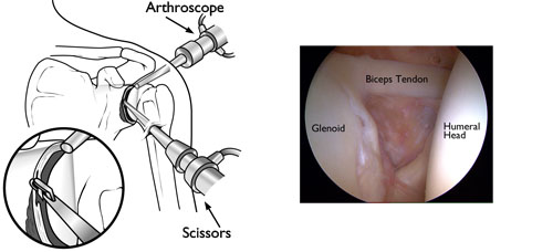
(Left) During arthroscopy, your surgeon inserts the arthroscope and small instruments into your shoulder joint. (Right) An arthroscopic view of the inside of the shoulder joint.
Once the problem is clearly identified, I will insert other small instruments through separate incisions to repair it. Specialized instruments are used for tasks like shaving, cutting, grasping, suture passing, and knot tying. In many cases, special devices are used to anchor stitches into bone.
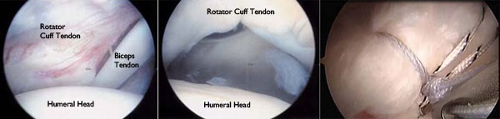
(Left) An arthroscopic view of a healthy shoulder joint. (Center) In this image of a rotator cuff tear, a large gap can be seen between the edge of the rotator cuff tendon and the humeral head. (Right) The tendon has been re-attached to the humeral head with sutures..
How would be Recovery Like?
After surgery, you will stay in the recovery room for 1 to 2 hours before being shifted to room. Nurses will monitor your responsiveness and provide pain medication.
Tell me something about Rehabilitation
Rehabilitation plays an important role in getting you back to your daily activities. An exercise program will help you regain shoulder strength and motion. I will develop a rehabilitation plan based on the surgical procedures you required.
If you have had a more complicated surgical repair, I may recommend a physical therapist to supervise your exercise program.
It is important that you make a strong effort at rehabilitation in order for your surgery to succeed.
What can be the possible Complications?
Most patients do not experience complications from shoulder arthroscopy. As with any surgery, however, there are some risks. These are usually minor and treatable. Potential problems with arthroscopy include infection, excessive bleeding, blood clots, and damage to blood vessels or nerves.
I will discuss the possible complications with you before your operation.